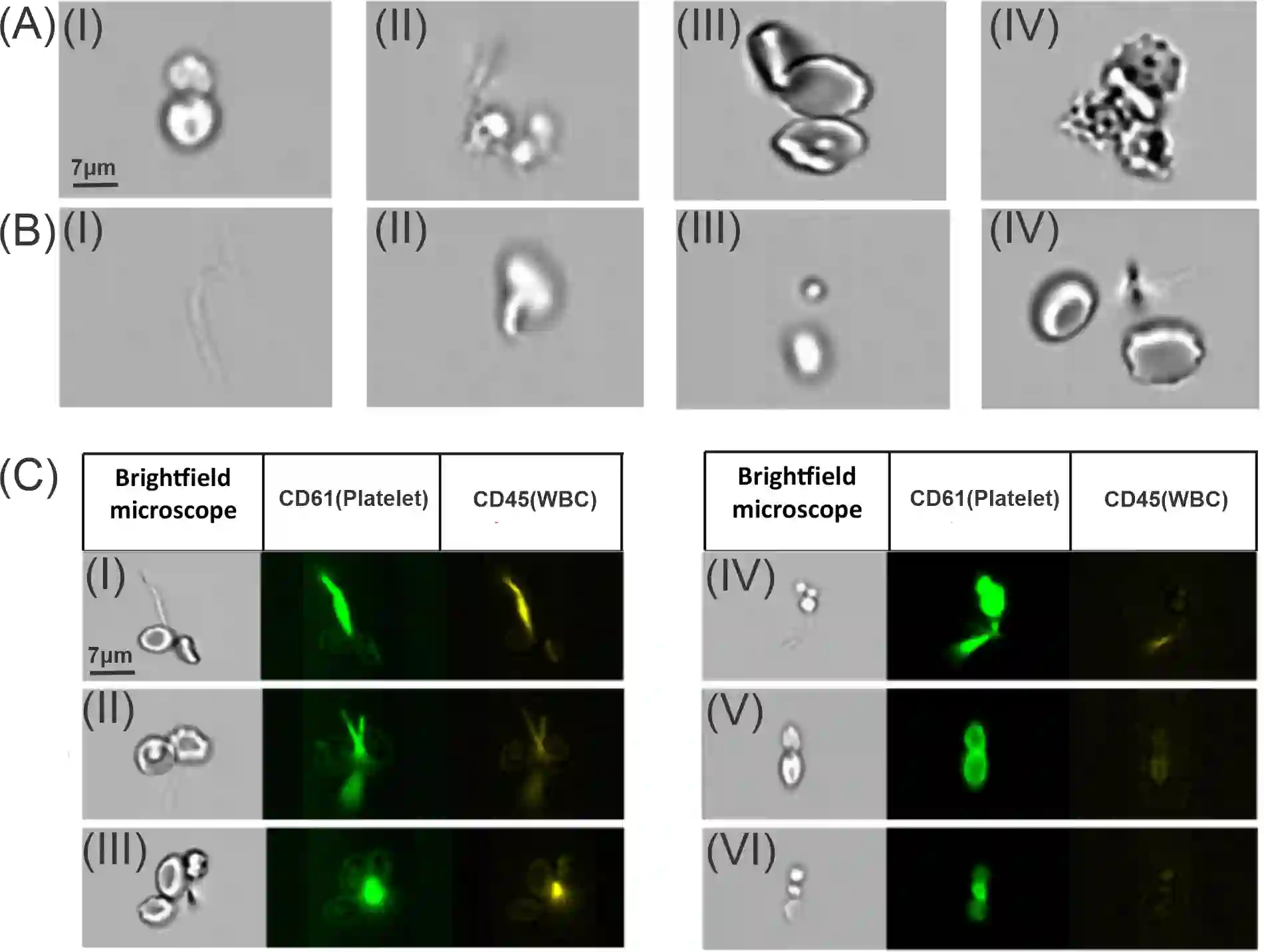
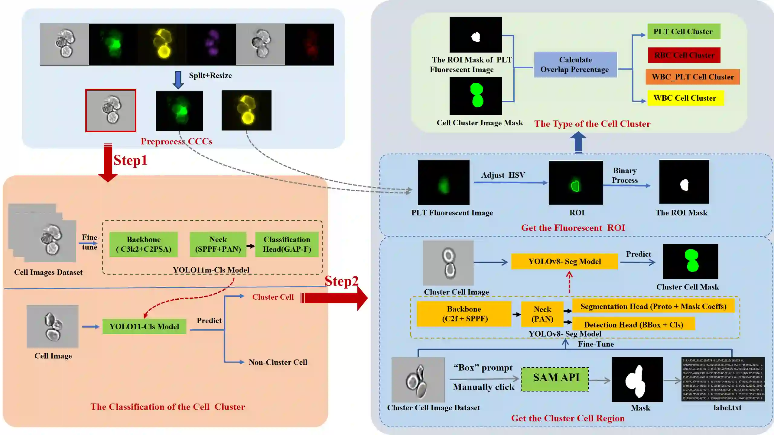
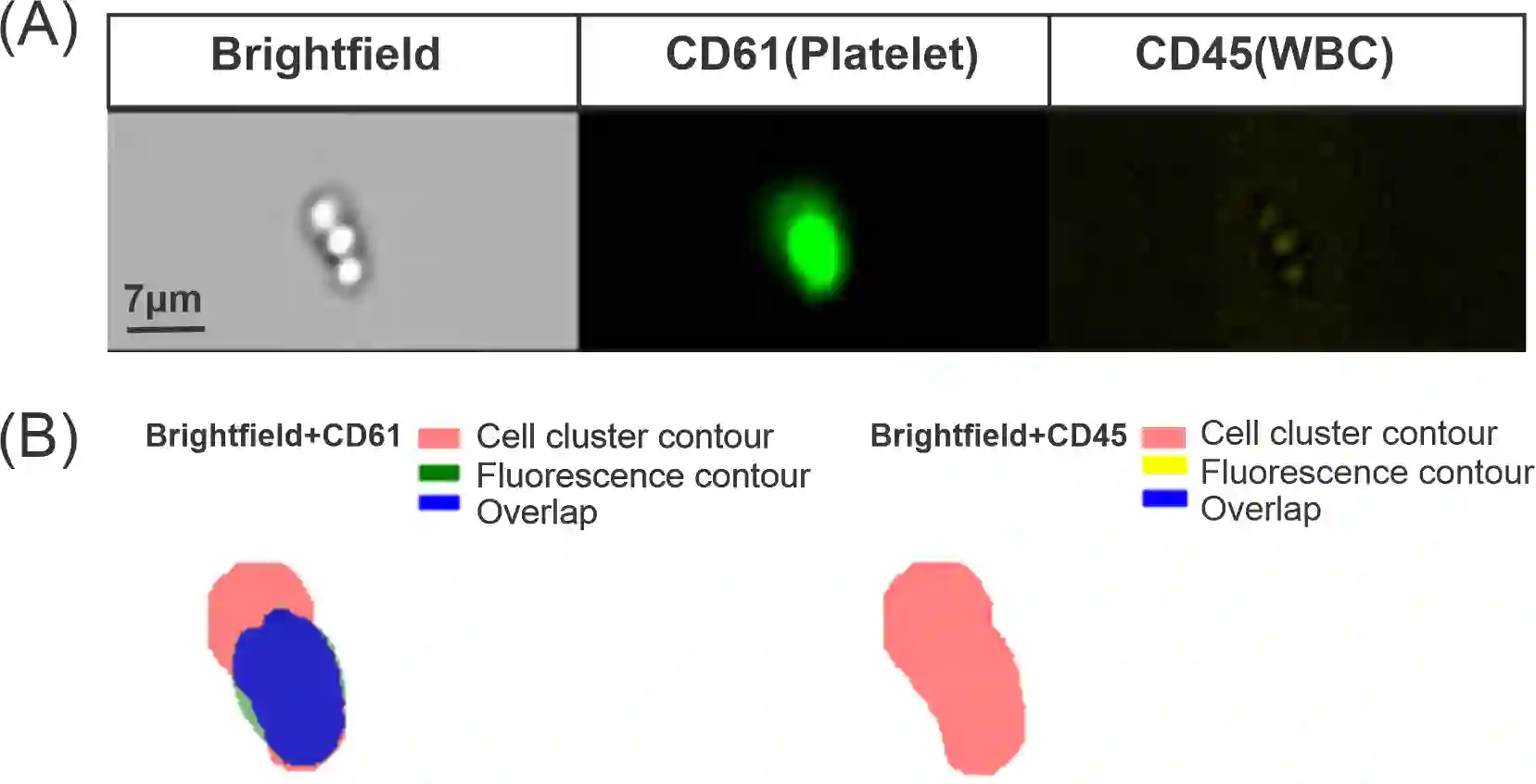
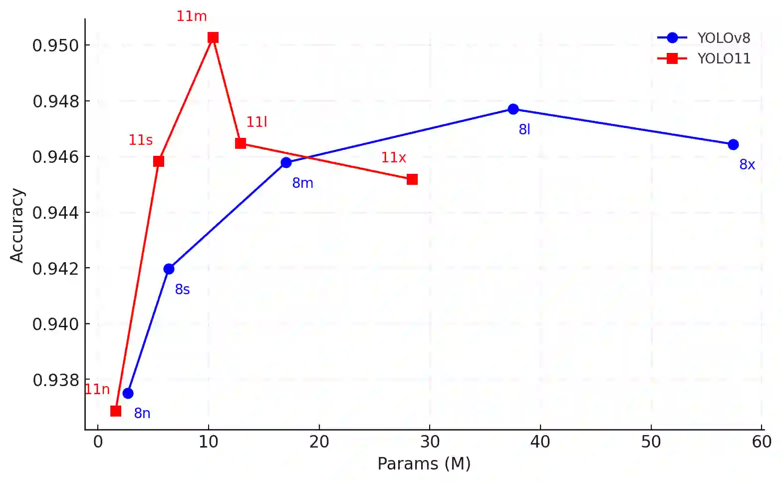
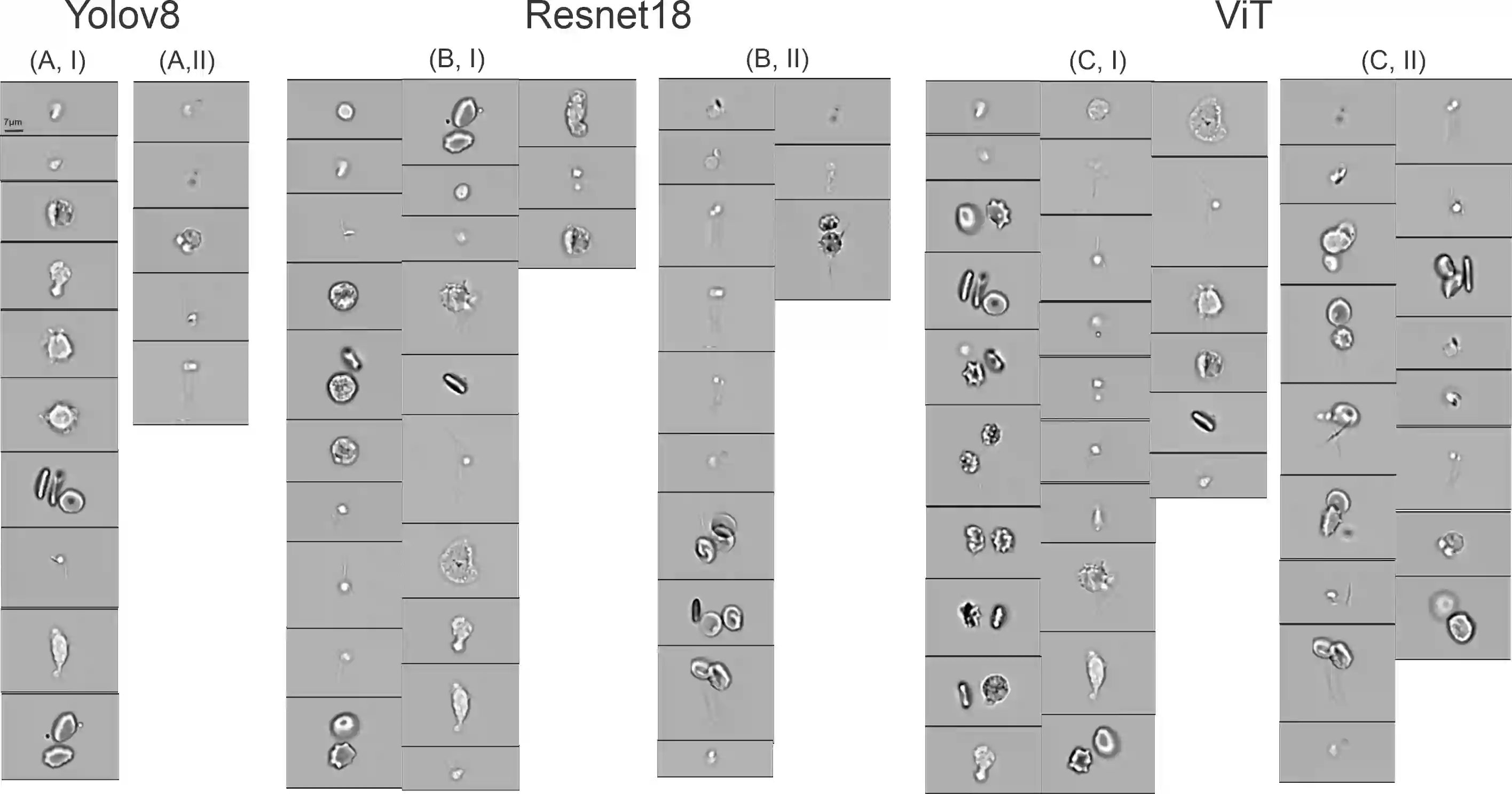
Circulating blood cell clusters (CCCs) containing red blood cells (RBCs), white blood cells(WBCs), and platelets are significant biomarkers linked to conditions like thrombosis, infection, and inflammation. Flow cytometry, paired with fluorescence staining, is commonly used to analyze these cell clusters, revealing cell morphology and protein profiles. While computational approaches based on machine learning have advanced the automatic analysis of single-cell flow cytometry images, there is a lack of effort to build tools to automatically analyze images containing CCCs. Unlike single cells, cell clusters often exhibit irregular shapes and sizes. In addition, these cell clusters often consist of heterogeneous cell types, which require multi-channel staining to identify the specific cell types within the clusters. This study introduces a new computational framework for analyzing CCC images and identifying cell types within clusters. Our framework uses a two-step analysis strategy. First, it categorizes images into cell cluster and non-cluster groups by fine-tuning the You Only Look Once(YOLOv11) model, which outperforms traditional convolutional neural networks (CNNs), Vision Transformers (ViT). Then, it identifies cell types by overlaying cluster contours with regions from multi-channel fluorescence stains, enhancing accuracy despite cell debris and staining artifacts. This approach achieved over 95% accuracy in both cluster classification and phenotype identification. In summary, our automated framework effectively analyzes CCC images from flow cytometry, leveraging both bright-field and fluorescence data. Initially tested on blood cells, it holds potential for broader applications, such as analyzing immune and tumor cell clusters, supporting cellular research across various diseases.
翻译:循环血细胞簇(CCCs)包含红细胞(RBCs)、白细胞(WBCs)和血小板,是与血栓形成、感染和炎症等病症相关的重要生物标志物。流式细胞术结合荧光染色常用于分析这些细胞簇,以揭示细胞形态和蛋白质谱。虽然基于机器学习的计算方法已推进了单细胞流式细胞图像的自动分析,但目前仍缺乏用于自动分析含CCCs图像的工具。与单细胞不同,细胞簇通常呈现不规则的形状和尺寸。此外,这些细胞簇常由异质性细胞类型组成,需要多通道染色来识别簇内的特定细胞类型。本研究提出了一种新的计算框架,用于分析CCC图像并识别簇内的细胞类型。我们的框架采用两步分析策略。首先,通过微调You Only Look Once(YOLOv11)模型将图像分类为细胞簇与非簇组,该模型性能优于传统的卷积神经网络(CNNs)和Vision Transformers(ViT)。随后,通过将细胞簇轮廓与多通道荧光染色区域叠加来识别细胞类型,从而在存在细胞碎片和染色伪影的情况下提高准确性。该方法在细胞簇分类和表型识别方面均达到了95%以上的准确率。总之,我们的自动化框架有效利用了明场和荧光数据,能够高效分析流式细胞术产生的CCC图像。该框架最初在血细胞上进行了测试,并具有更广泛应用的潜力,例如分析免疫细胞和肿瘤细胞簇,从而支持针对多种疾病的细胞研究。