
The electron microscope (EM) remains the predominant technique for elucidating intricate details of the animal nervous system at the nanometer scale. However, accurately reconstructing the complex morphology of axons and myelin sheaths poses a significant challenge. Furthermore, the absence of publicly available, large-scale EM datasets encompassing complete cross sections of the corpus callosum, with dense ground truth segmentation for axons and myelin sheaths, hinders the advancement and evaluation of holistic corpus callosum reconstructions. To surmount these obstacles, we introduce the AxonCallosumEM dataset, comprising a 1.83 times 5.76mm EM image captured from the corpus callosum of the Rett Syndrome (RTT) mouse model, which entail extensive axon bundles. We meticulously proofread over 600,000 patches at a resolution of 1024 times 1024, thus providing a comprehensive ground truth for myelinated axons and myelin sheaths. Additionally, we extensively annotated three distinct regions within the dataset for the purposes of training, testing, and validation. Utilizing this dataset, we develop a fine-tuning methodology that adapts Segment Anything Model (SAM) to EM images segmentation tasks, called EM-SAM, enabling outperforms other state-of-the-art methods. Furthermore, we present the evaluation results of EM-SAM as a baseline.
翻译:电子显微镜仍然是揭示动物神经系统纳米尺度细微结构的主要技术手段。然而,精确重建轴突和髓鞘的复杂形态仍是一项重大挑战。此外,缺乏包含完整胼胝体横截面的公开大规模电镜数据集(附有密集标注的轴突和髓鞘真实分割结果),阻碍了整体胼胝体重建的进展与评估。为克服这些障碍,我们引入了AxonCallosumEM数据集,该数据集包含从瑞特综合征小鼠模型胼胝体采集的1.83×5.76毫米电镜图像,其中包含大量轴突束。我们仔细校对超过60万张1024×1024分辨率的图像块,从而提供了有髓轴突和髓鞘的全面真实标注。此外,我们在数据集中对三个不同区域进行了广泛标注,用于训练、测试和验证。利用该数据集,我们开发了一种微调方法,将Segment Anything Model适配至电镜图像分割任务(称为EM-SAM),使其性能超越其他现有最优方法。同时,我们给出了EM-SAM作为基线的评估结果。
相关内容
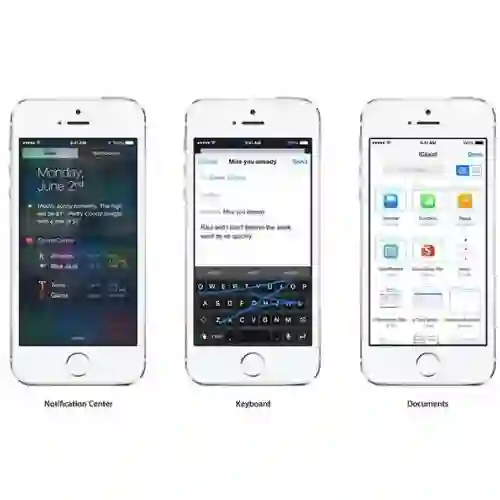
- Today (iOS and OS X): widgets for the Today view of Notification Center
- Share (iOS and OS X): post content to web services or share content with others
- Actions (iOS and OS X): app extensions to view or manipulate inside another app
- Photo Editing (iOS): edit a photo or video in Apple's Photos app with extensions from a third-party apps
- Finder Sync (OS X): remote file storage in the Finder with support for Finder content annotation
- Storage Provider (iOS): an interface between files inside an app and other apps on a user's device
- Custom Keyboard (iOS): system-wide alternative keyboards
Source: iOS 8 Extensions: Apple’s Plan for a Powerful App Ecosystem


