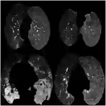
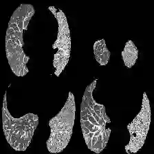
The pandemic resulted in vast repositories of unstructured data, including radiology reports, due to increased medical examinations. Previous research on automated diagnosis of COVID-19 primarily focuses on X-ray images, despite their lower precision compared to computed tomography (CT) scans. In this work, we leverage unstructured data from a hospital and harness the fine-grained details offered by CT scans to perform zero-shot multi-label classification based on contrastive visual language learning. In collaboration with human experts, we investigate the effectiveness of multiple zero-shot models that aid radiologists in detecting pulmonary embolisms and identifying intricate lung details like ground glass opacities and consolidations. Our empirical analysis provides an overview of the possible solutions to target such fine-grained tasks, so far overlooked in the medical multimodal pretraining literature. Our investigation promises future advancements in the medical image analysis community by addressing some challenges associated with unstructured data and fine-grained multi-label classification.
翻译:大规模医疗检查导致疫情催生了大量非结构化数据,包括放射学报告。尽管计算机断层扫描(CT)的精度高于X光影像,以往COVID-19自动诊断研究仍主要聚焦于X光图像。本研究利用医院的非结构化数据,结合CT扫描提供的精细细节,基于对比视觉语言学习执行零样本多标签分类。我们与人类专家合作,评估了多种零样本模型在辅助放射科医生检测肺栓塞、识别磨玻璃影和实变等复杂肺部细节方面的有效性。实证分析概述了针对这些迄今在医学多模态预训练文献中鲜少涉及的细粒度任务的可能解决方案。本研究通过应对非结构化数据与细粒度多标签分类的挑战,为医学图像分析领域的未来发展提供了前景。