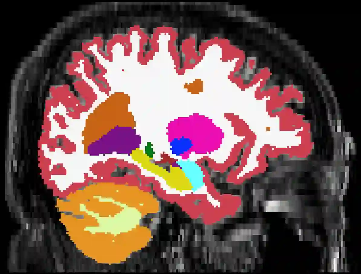
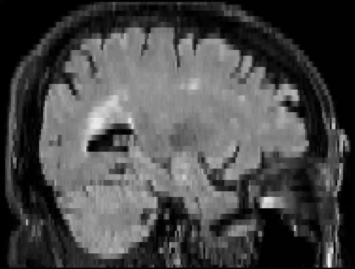
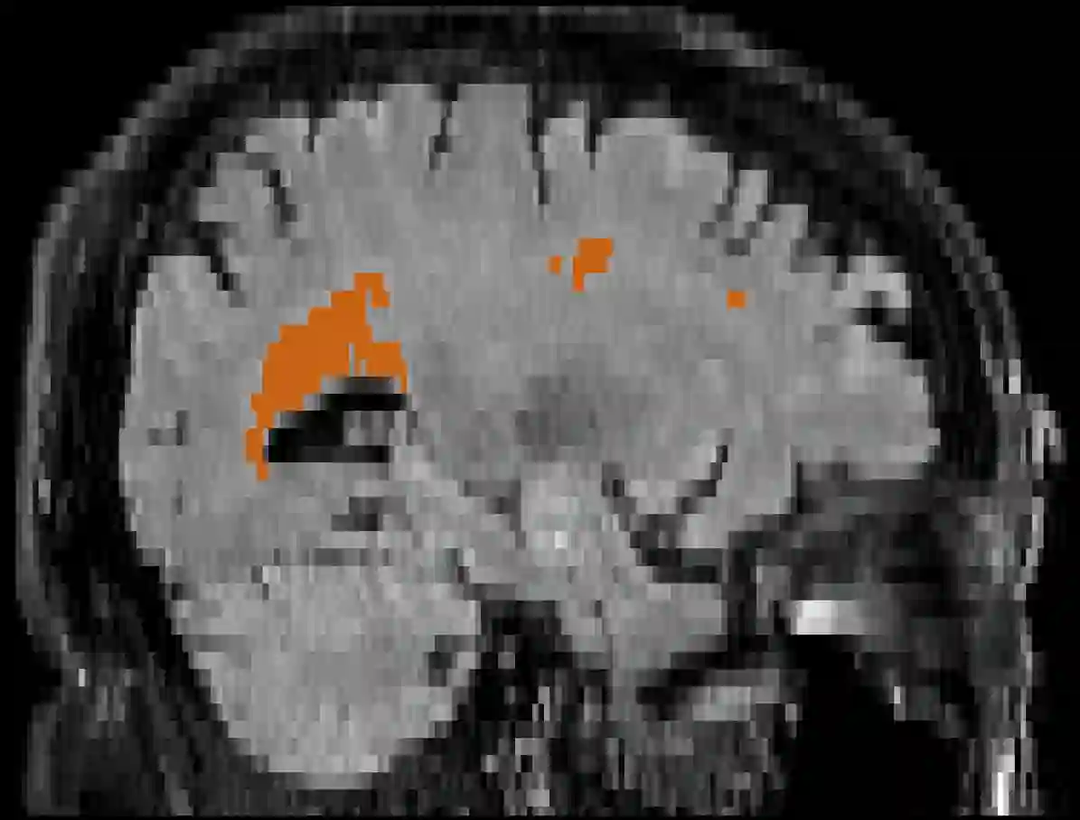
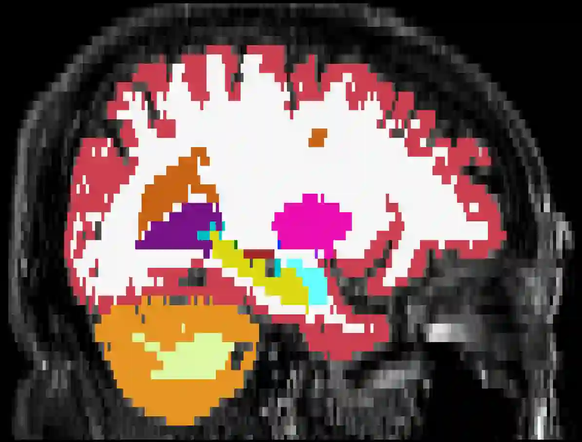
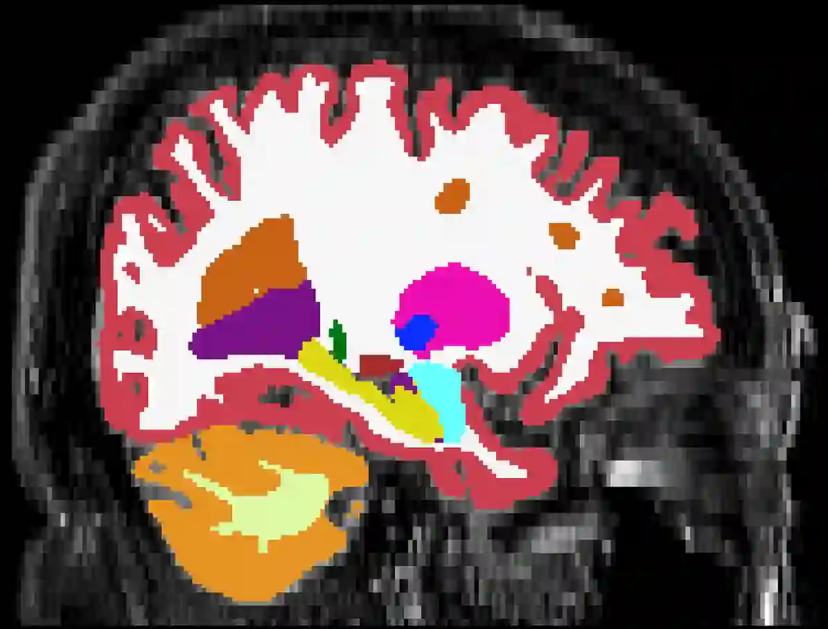
Brain atrophy and white matter hyperintensity (WMH) are critical neuroimaging features for ascertaining brain injury in cerebrovascular disease and multiple sclerosis. Automated segmentation and quantification is desirable but existing methods require high-resolution MRI with good signal-to-noise ratio (SNR). This precludes application to clinical and low-field portable MRI (pMRI) scans, thus hampering large-scale tracking of atrophy and WMH progression, especially in underserved areas where pMRI has huge potential. Here we present a method that segments white matter hyperintensity and 36 brain regions from scans of any resolution and contrast (including pMRI) without retraining. We show results on six public datasets and on a private dataset with paired high- and low-field scans (3T and 64mT), where we attain strong correlation between the WMH ($\rho$=.85) and hippocampal volumes (r=.89) estimated at both fields. Our method is publicly available as part of FreeSurfer, at: http://surfer.nmr.mgh.harvard.edu/fswiki/WMH-SynthSeg.
翻译:脑萎缩和白质高信号是脑血管疾病和多发性硬化中评估脑损伤的关键神经影像学特征。自动分割与量化方法虽然理想,但现有技术需要具有良好信噪比的高分辨率MRI,从而无法应用于临床和低场便携式MRI扫描,阻碍了脑萎缩和WMH进展的大规模追踪,尤其是在pMRI具有巨大潜力的医疗资源匮乏地区。本文提出一种无需重新训练即可从任意分辨率和对比度(包括pMRI)的扫描中分割白质高信号及36个脑区的方法。我们在六个公共数据集和包含配对高/低场扫描(3T与64mT)的私有数据集上展示了结果,两个场强下估计的WMH($\rho$=.85)和海马体积(r=.89)均呈现强相关性。本方法已作为FreeSurfer的一部分公开发布,网址:http://surfer.nmr.mgh.harvard.edu/fswiki/WMH-SynthSeg。