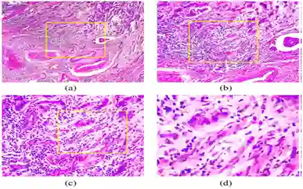
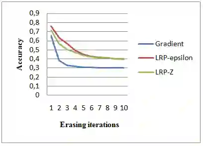
Computer aided detection and diagnosis systems based on deep learning have shown promising performance in breast cancer detection. However, there are cases where the obtained results lack justification. In this study, our objective is to highlight the regions of interest used by a convolutional neural network (CNN) for classifying histological images as benign or malignant. We compare these regions with the regions identified by pathologists. To achieve this, we employed the VGG19 architecture and tested three visualization methods: Gradient, LRP Z, and LRP Epsilon. Additionally, we experimented with three pixel selection methods: Bins, K-means, and MeanShift. Based on the results obtained, the Gradient visualization method and the MeanShift selection method yielded satisfactory outcomes for visualizing the images.
翻译:基于深度学习的计算机辅助检测与诊断系统在乳腺癌检测中展现出良好性能。然而,部分结果缺乏可解释性。本研究旨在突出卷积神经网络(CNN)在区分良恶性组织学图像时关注的感兴趣区域,并将这些区域与病理学家标注的区域进行对比。为此,我们采用VGG19架构,测试了三种可视化方法:Gradient、LRP Z和LRP Epsilon,并实验了三种像素选择方法:Bins、K-means和MeanShift。实验结果表明,Gradient可视化方法与MeanShift选择方法在图像可视化方面取得了令人满意的效果。