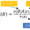
Background and purpose: The unanticipated detection by magnetic resonance imaging (MRI) in the brain of asymptomatic subjects of white matter lesions suggestive of multiple sclerosis (MS) has been named radiologically isolated syndrome (RIS). As the difference between early MS [i.e. clinically isolated syndrome (CIS)] and RIS is the occurrence of a clinical event, it is logical to improve detection of the subclinical form without interfering with MRI as there are radiological diagnostic criteria for that. Our objective was to use machine-learning classification methods to identify morphometric measures that help to discriminate patients with RIS from those with CIS. Methods: We used a multimodal 3-T MRI approach by combining MRI biomarkers (cortical thickness, cortical and subcortical grey matter volume, and white matter integrity) of a cohort of 17 patients with RIS and 17 patients with CIS for single-subject level classification. Results: The best proposed models to predict the diagnosis of CIS and RIS were based on the Naive Bayes, Bagging and Multilayer Perceptron classifiers using only three features: the left rostral middle frontal gyrus volume and the fractional anisotropy values in the right amygdala and right lingual gyrus. The Naive Bayes obtained the highest accuracy [overall classification, 0.765; area under the receiver operating characteristic (AUROC), 0.782]. Conclusions: A machine-learning approach applied to multimodal MRI data may differentiate between the earliest clinical expressions of MS (CIS and RIS) with an accuracy of 78%. Keywords: Bagging; Multilayer Perceptron; Naive Bayes classifier; clinically isolated syndrome; diffusion tensor imaging; machine-learning; magnetic resonance imaging; multiple sclerosis; radiologically isolated syndrome.
翻译:背景与目的:在无症状受试者脑部磁共振成像(MRI)中意外检测到提示多发性硬化(MS)的白质病变,被称为放射学孤立综合征(RIS)。由于早期MS(即临床孤立综合征,CIS)与RIS的差异在于临床事件的发生,因此在不干扰MRI的情况下,利用放射学诊断标准来改进对亚临床形式的识别具有逻辑性。我们的目标是使用机器学习分类方法识别有助于区分RIS患者与CIS患者的形态测量指标。方法:我们采用多模态3T MRI方法,結合MRI生物标志物(皮层厚度、皮层及皮层下灰质体积、白质完整性),对17例RIS患者和17例CIS患者进行单受试者水平分类。结果:预测CIS与RIS诊断的最佳模型基于朴素贝叶斯、Bagging和多层感知器分类器,仅使用三个特征:左侧额中回喙部体积、右侧杏仁核及右侧舌回的分数各向异性值。朴素贝叶斯获得了最高准确率(总体分类为0.765;受试者工作特征曲线下面积AUROC为0.782)。结论:将机器学习方法应用于多模态MRI数据,能以78%的准确率区分MS的最早期临床表现(CIS与RIS)。关键词:Bagging;多层感知器;朴素贝叶斯分类器;临床孤立综合征;弥散张量成像;机器学习;磁共振成像;多发性硬化;放射学孤立综合征。