

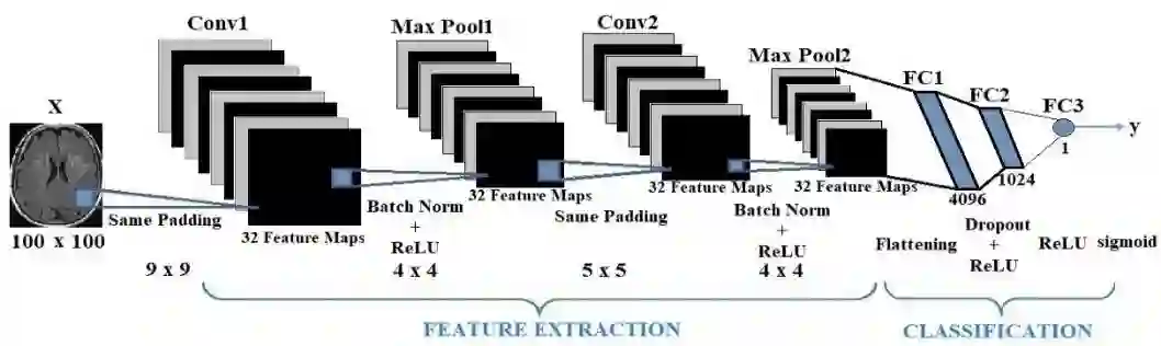
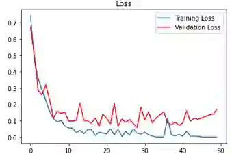
The automated detection of cancerous tumors has attracted interest mainly during the last decade, due to the necessity of early and efficient diagnosis that will lead to the most effective possible treatment of the impending risk. Several machine learning and artificial intelligence methodologies has been employed aiming to provide trustworthy helping tools that will contribute efficiently to this attempt. In this article, we present a low-complexity convolutional neural network architecture for tumor classification enhanced by a robust image augmentation methodology. The effectiveness of the presented deep learning model has been investigated based on 3 datasets containing brain, kidney and lung images, showing remarkable diagnostic efficiency with classification accuracies of 99.33%, 100% and 99.7% for the 3 datasets respectively. The impact of the augmentation preprocessing step has also been extensively examined using 4 evaluation measures. The proposed low-complexity scheme, in contrast to other models in the literature, renders our model quite robust to cases of overfitting that typically accompany small datasets frequently encountered in medical classification challenges. Finally, the model can be easily re-trained in case additional volume images are included, as its simplistic architecture does not impose a significant computational burden.
翻译:癌性肿瘤的自动检测在过去十年间引起了广泛关注,这源于早期有效诊断的需求,以便对潜在风险采取最有效的治疗。多种机器学习和人工智能方法已被用于提供值得信赖的辅助工具,以有效推动这一努力。本文提出了一种低复杂度的卷积神经网络架构,用于肿瘤分类,并通过鲁棒的图像增强方法进行了优化。基于包含脑部、肾脏和肺部图像的三个数据集对所提深度学习模型的有效性进行了研究,结果显示其诊断效率显著,三个数据集的分类准确率分别达到99.33%、100%和99.7%。同时,还使用了四种评估指标对增强预处理步骤的影响进行了广泛检验。与文献中的其他模型相比,所提出的低复杂度方案使我们的模型对医学分类挑战中常见的小数据集容易出现的过拟合问题具有较强的鲁棒性。最后,由于模型架构简单,不会带来显著的计算负担,因此在新增体积图像时可以轻松重新训练。




相关内容





 微信扫码咨询专知VIP会员