

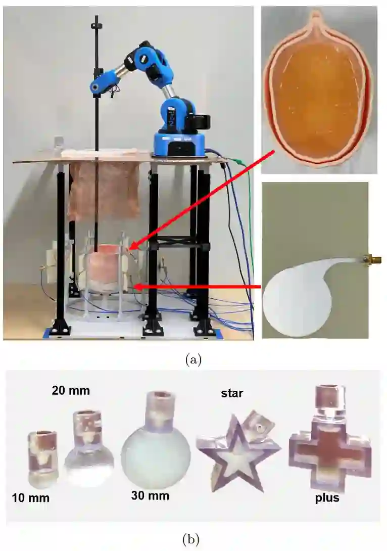
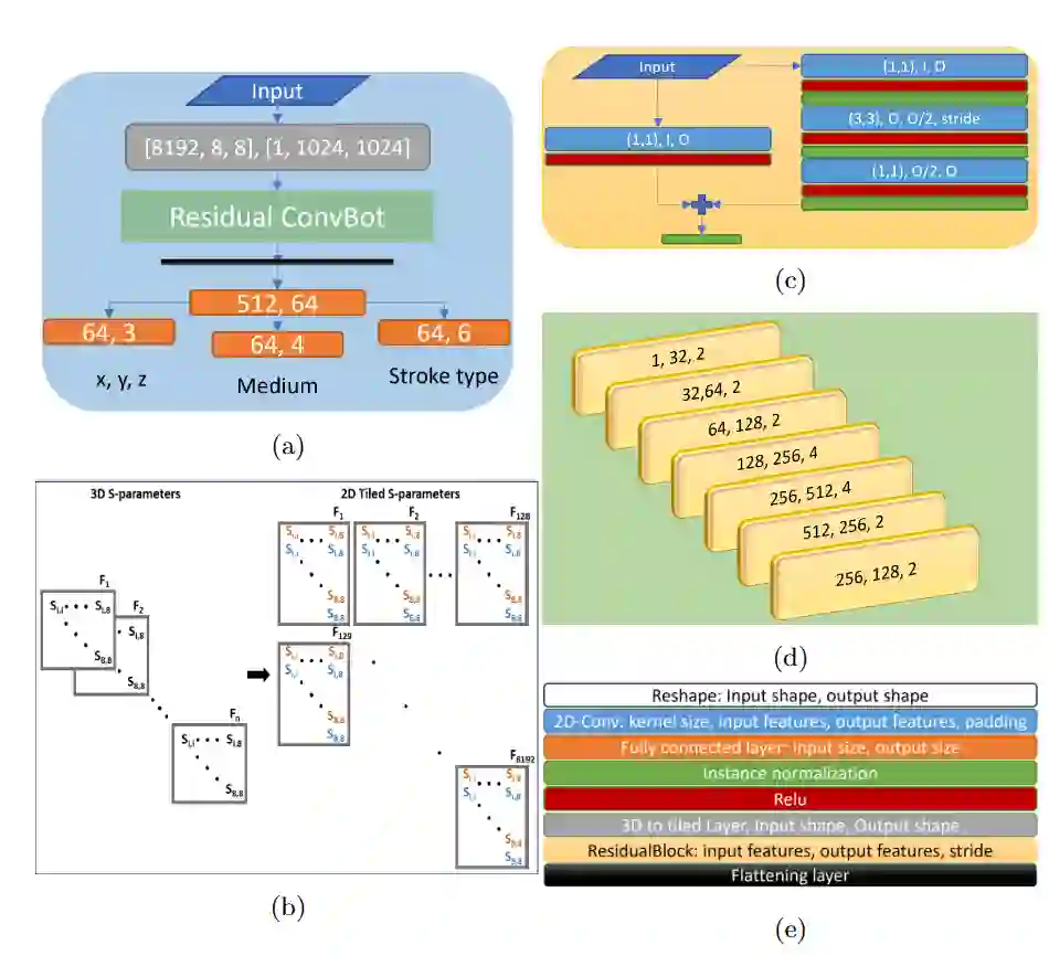
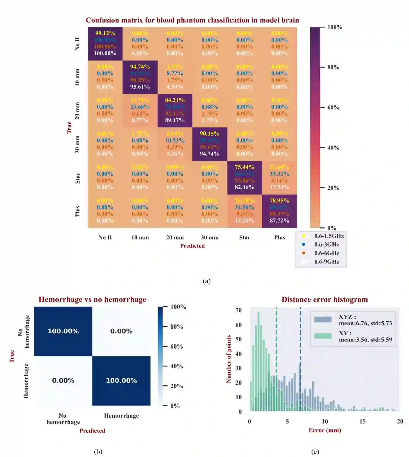
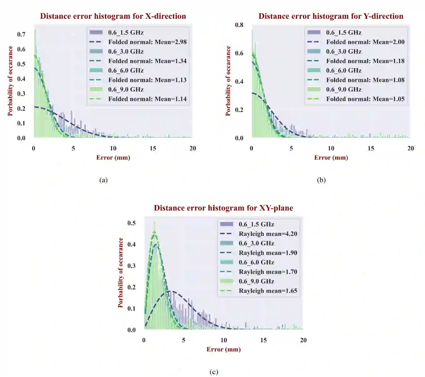
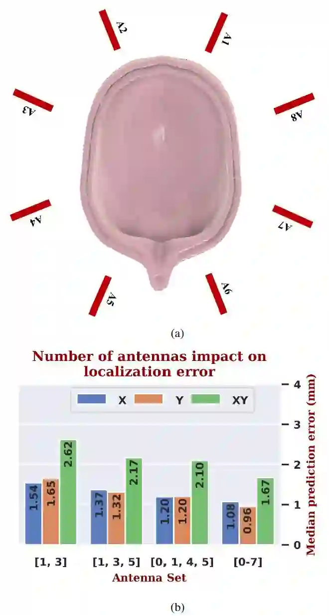
Stroke is a leading cause of mortality and disability. Emergent diagnosis and intervention are critical, and predicated upon initial brain imaging; however, existing clinical imaging modalities are generally costly, immobile, and demand highly specialized operation and interpretation. Low-energy microwaves have been explored as low-cost, small form factor, fast, and safe probes of tissue dielectric properties, with both imaging and diagnostic potential. Nevertheless, challenges inherent to microwave reconstruction have impeded progress, hence microwave imaging (MWI) remains an elusive scientific aim. Herein, we introduce a dedicated experimental framework comprising a robotic navigation system to translate blood-mimicking phantoms within an anatomically realistic human head model. An 8-element ultra-wideband (UWB) array of modified antipodal Vivaldi antennas was developed and driven by a two-port vector network analyzer spanning 0.6-9.0 GHz at an operating power of 1 mw. Complex scattering parameters were measured, and dielectric signatures of hemorrhage were learned using a dedicated deep neural network for prediction of hemorrhage classes and localization. An overall sensitivity and specificity for detection >0.99 was observed, with Rayliegh mean localization error of 1.65 mm. The study establishes the feasibility of a robust experimental model and deep learning solution for UWB microwave stroke detection.
翻译:脑卒中是导致死亡和残疾的主要原因。紧急诊断和干预至关重要,且依赖于初始脑部成像;然而,现有的临床成像方式通常成本高昂、不便移动,且需要高度专业化的操作和解读。低能量微波因其低成本、小体积、快速且安全探查组织介电特性的优势,兼具成像和诊断潜力,已被广泛研究。然而,微波重建固有的挑战阻碍了其发展,使得微波成像(MWI)仍是一个难以实现的科学目标。本文介绍了一套专用实验框架,包括一个机器人导航系统,用于在解剖学逼真的人体头部模型中移动模拟血液的体模。我们开发了一个8单元超宽带(UWB)改进型对跖Vivaldi天线阵列,由双端口矢量网络分析仪驱动,工作频率范围为0.6-9.0 GHz,工作功率为1 mW。测量了复散射参数,并利用专用深度神经网络学习出血的介电特征,以预测出血类别和定位。观察到的整体检测灵敏度和特异性均大于0.99,瑞利平均定位误差为1.65毫米。本研究证实了用于UWB脑卒中检测的鲁棒实验模型和深度学习解决方案的可行性。




相关内容




 微信扫码咨询专知VIP会员