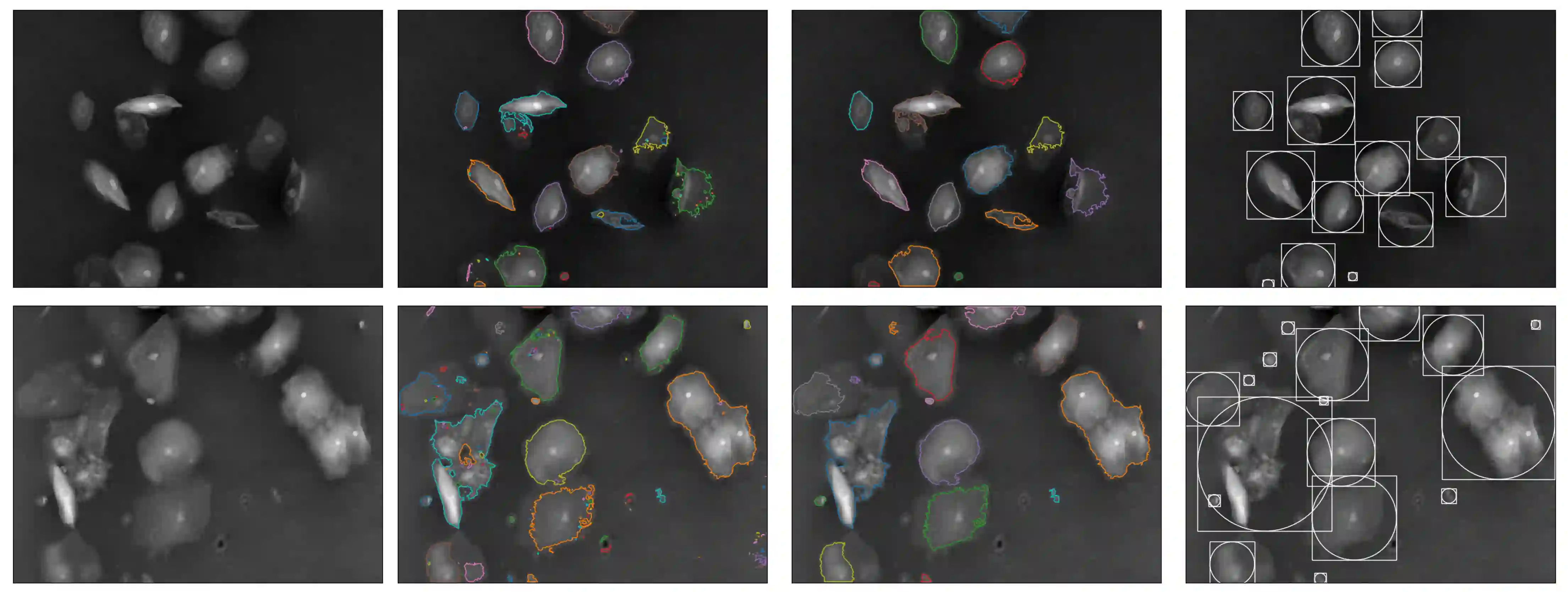
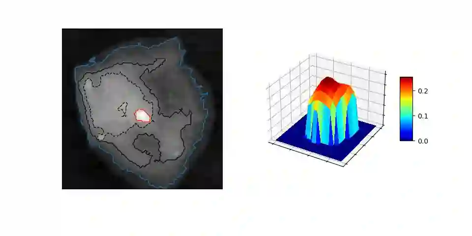
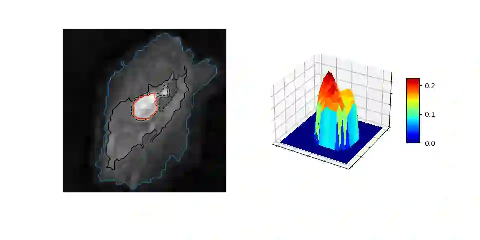
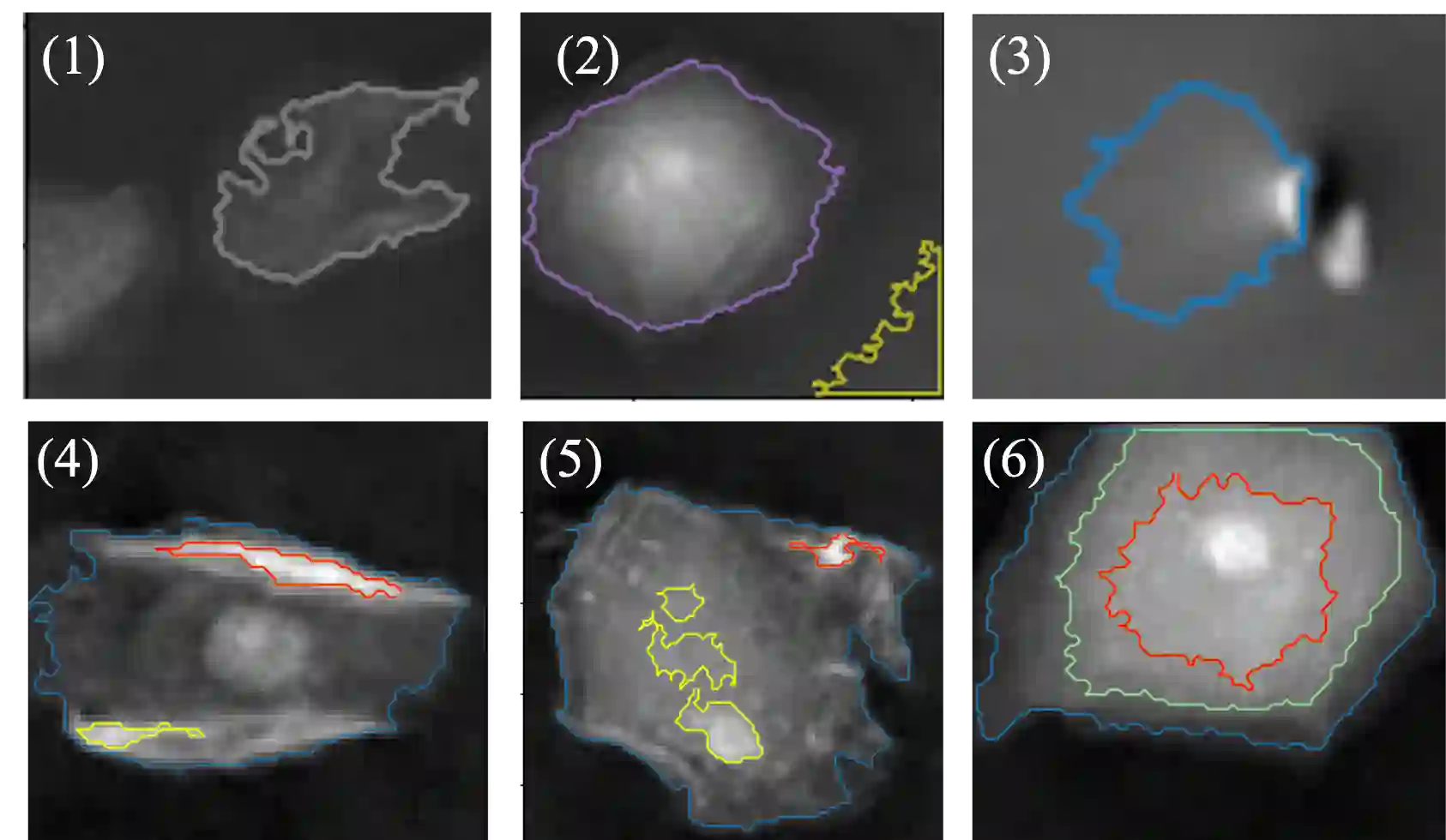
In the effort to aid cytologic diagnostics by establishing automatic single cell screening using high throughput digital holographic microscopy for clinical studies thousands of images and millions of cells are captured. The bottleneck lies in an automatic, fast, and unsupervised segmentation technique that does not limit the types of cells which might occur. We propose an unsupervised multistage method that segments correctly without confusing noise or reflections with cells and without missing cells that also includes the detection of relevant inner structures, especially the cell nucleus in the unstained cell. In an effort to make the information reasonable and interpretable for cytopathologists, we also introduce new cytoplasmic and nuclear features of potential help for cytologic diagnoses which exploit the quantitative phase information inherent to the measurement scheme. We show that the segmentation provides consistently good results over many experiments on patient samples in a reasonable per cell analysis time.
翻译:为通过建立基于高通量数字全息显微术的自动单细胞筛查辅助细胞学诊断,临床研究中需捕获数千张图像及数百万个细胞。当前技术瓶颈在于缺乏一种自动、快速且无监督的分割方法——该方法需不限制可能出现的细胞类型,既能准确分割细胞而不将噪声或反射误判为细胞,亦不遗漏细胞,同时还能检测相关内部结构(尤其是未染色细胞中的细胞核)。为使信息对细胞病理学家合理且可解释,本文还引入了两类利用测量方案固有定量相位信息的新型细胞质与细胞核特征,有望辅助细胞学诊断。实验表明,该分割方法在患者样本的多次实验中均能稳定取得良好结果,且单细胞分析时间合理。