

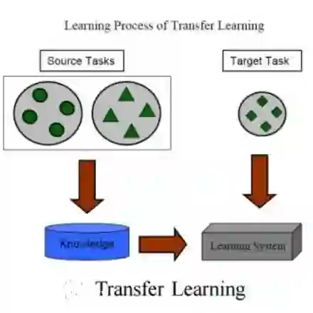
A major challenge in stroke research and stroke recovery predictions is the determination of a stroke lesion's extent and its impact on relevant brain systems. Manual segmentation of stroke lesions from 3D magnetic resonance (MR) imaging volumes, the current gold standard, is not only very time-consuming, but its accuracy highly depends on the operator's experience. As a result, there is a need for a fully automated segmentation method that can efficiently and objectively measure lesion extent and the impact of each lesion to predict impairment and recovery potential which might be beneficial for clinical, translational, and research settings. We have implemented and tested a fully automatic method for stroke lesion segmentation which was developed using eight different 2D-model architectures trained via transfer learning (TL) and mixed data approaches. Additionally, the final prediction was made using a novel ensemble method involving stacking and agreement window. Our novel method was evaluated in a novel in-house dataset containing 22 T1w brain MR images, which were challenging in various perspectives, but mostly because they included T1w MR images from the subacute (which typically less well defined T1 lesions) and chronic stroke phase (which typically means well defined T1-lesions). Cross-validation results indicate that our new method can efficiently and automatically segment lesions fast and with high accuracy compared to ground truth. In addition to segmentation, we provide lesion volume and weighted lesion load of relevant brain systems based on the lesions' overlap with a canonical structural motor system that stretches from the cortical motor region to the lowest end of the brain stem.
翻译:脑卒中研究与康复预测中的一大挑战在于确定卒中病灶的范围及其对相关脑系统的影响。当前金标准——从3D磁共振成像体积中手动分割卒中病灶——不仅极为耗时,其准确性还高度依赖于操作者的经验。因此,亟需一种全自动分割方法,能够高效且客观地测量病灶范围及每个病灶的影响,从而预测功能障碍与康复潜力,这可能对临床、转化医学及研究领域有所裨益。我们实现并测试了一种全自动卒中病灶分割方法,该方法是基于八种不同的2D模型架构开发的,这些架构通过迁移学习和混合数据方法进行训练。此外,最终预测采用了一种新颖的集成方法,该方法涉及堆叠与一致性窗口。我们的新方法在一个包含22个T1加权脑部MRI图像的新内部数据集上进行了评估,这些图像在多方面具有挑战性,但主要难点在于它们包含了亚急性期(通常T1病灶定义不清)和慢性期(通常T1病灶定义清晰)的T1加权MRI图像。交叉验证结果表明,与金标准相比,我们的新方法能够快速高效地自动分割病灶,且准确性较高。除分割外,我们还根据病灶与一个从皮层运动区延伸至脑干最底端的典型结构性运动系统的重叠情况,提供了相关脑系统的病灶体积与加权病灶负荷。




相关内容




 微信扫码咨询专知VIP会员