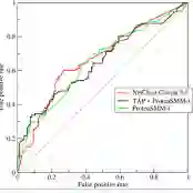
Background: Rim+ lesions in multiple sclerosis (MS), detectable via Quantitative Susceptibility Mapping (QSM), correlate with increased disability. Existing literature lacks quantitative analysis of these lesions. We introduce RimSet for quantitative identification and characterization of rim+ lesions on QSM. Methods: RimSet combines RimSeg, an unsupervised segmentation method using level-set methodology, and radiomic measurements with Local Binary Pattern texture descriptors. We validated RimSet using simulated QSM images and an in vivo dataset of 172 MS subjects with 177 rim+ and 3986 rim-lesions. Results: RimSeg achieved a 78.7% Dice score against the ground truth, with challenges in partial rim lesions. RimSet detected rim+ lesions with a partial ROC AUC of 0.808 and PR AUC of 0.737, surpassing existing methods. QSMRim-Net showed the lowest mean square error (0.85) and high correlation (0.91; 95% CI: 0.88, 0.93) with expert annotations at the subject level.
翻译:背景:多发性硬化(MS)中的Rim+病灶可通过定量磁敏感图(QSM)检测,并与功能残疾加重相关。现有文献缺乏对这些病灶的定量分析。我们提出RimSet方法,用于在QSM上定量识别并表征Rim+病灶。方法:RimSet结合了基于水平集方法的无监督分割算法RimSeg,以及采用局部二值模式纹理描述符的影像组学测量。我们通过模拟QSM图像和包含172名MS受试者(含177个Rim+病灶和3986个Rim−病灶)的体内数据集对RimSet进行验证。结果:与金标准相比,RimSeg的Dice系数达78.7%,但在处理部分边缘病灶时存在挑战。RimSet检测Rim+病灶的部分受试者工作特征曲线下面积(partial ROC AUC)为0.808,精确率-召回率曲线下面积(PR AUC)为0.737,优于现有方法。在受试者水平上,QSMRim-Net的均方误差最低(0.85),且与专家标注的相关性较高(0.91;95%置信区间:0.88,0.93)。