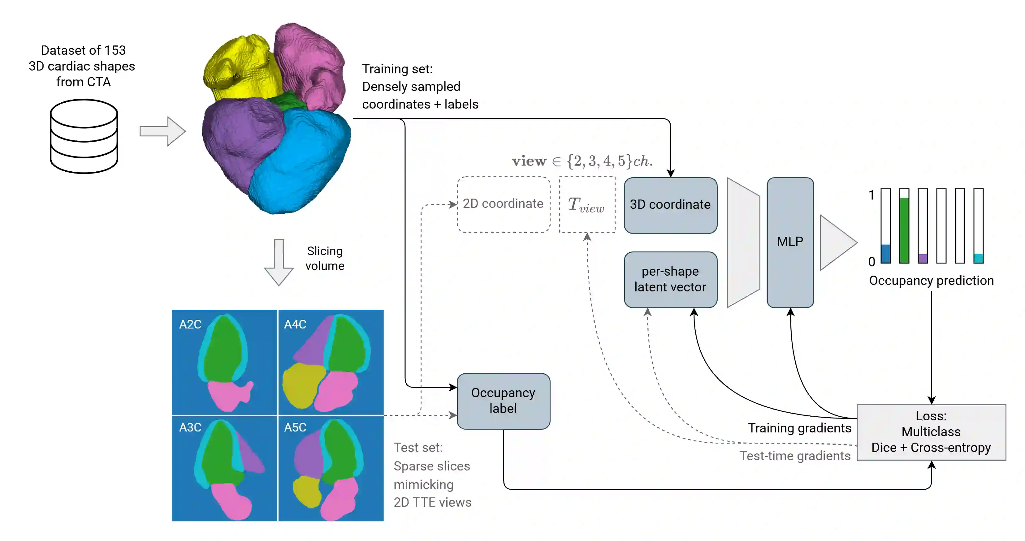
Accurate 3D representations of cardiac structures allow quantitative analysis of anatomy and function. In this work, we propose a method for reconstructing complete 3D cardiac shapes from segmentations of sparse planes in CT angiography (CTA) for application in 2D transthoracic echocardiography (TTE). Our method uses a neural implicit function to reconstruct the 3D shape of the cardiac chambers and left-ventricle myocardium from sparse CTA planes. To investigate the feasibility of achieving 3D reconstruction from 2D TTE, we select planes that mimic the standard apical 2D TTE views. During training, a multi-layer perceptron learns shape priors from 3D segmentations of the target structures in CTA. At test time, the network reconstructs 3D cardiac shapes from segmentations of TTE-mimicking CTA planes by jointly optimizing the latent code and the rigid transforms that map the observed planes into 3D space. For each heart, we simulate four realistic apical views, and we compare reconstructed multi-class volumes with the reference CTA volumes. On a held-out set of CTA segmentations, our approach achieves an average Dice coefficient of 0.86 $\pm$ 0.04 across all structures. Our method also achieves markedly lower volume errors than the clinical standard, Simpson's biplane rule: 4.88 $\pm$ 4.26 mL vs. 8.14 $\pm$ 6.04 mL, respectively, for the left ventricle; and 6.40 $\pm$ 7.37 mL vs. 37.76 $\pm$ 22.96 mL, respectively, for the left atrium. This suggests that our approach offers a viable route to more accurate 3D chamber quantification in 2D transthoracic echocardiography.
翻译:精确的三维心脏结构表征有助于实现解剖与功能的定量分析。本研究提出一种方法,能够从计算机断层扫描血管造影(CTA)的稀疏平面分割结果中重建完整的三维心脏形态,以应用于二维经胸超声心动图(TTE)。该方法采用神经隐式函数,从稀疏的CTA平面重建心脏各腔室及左心室心肌的三维形状。为探究从二维TTE实现三维重建的可行性,我们选取了模拟标准心尖二维TTE切面的平面。在训练阶段,多层感知机从CTA中目标结构的三维分割数据学习形状先验。在测试阶段,网络通过联合优化潜在编码与将观测平面映射至三维空间的刚性变换,从模拟TTE的CTA平面分割结果中重建三维心脏形状。针对每个心脏,我们模拟了四个真实的心尖切面,并将重建的多类别体积与参考CTA体积进行对比。在预留的CTA分割数据集上,本方法在所有结构上的平均Dice系数达到0.86 $\pm$ 0.04。与临床金标准Simpson双平面法相比,本方法的体积误差显著降低:左心室误差分别为4.88 $\pm$ 4.26 mL vs. 8.14 $\pm$ 6.04 mL;左心房误差分别为6.40 $\pm$ 7.37 mL vs. 37.76 $\pm$ 22.96 mL。这表明我们的方法为在二维经胸超声心动图中实现更精确的三维心腔量化提供了可行路径。