

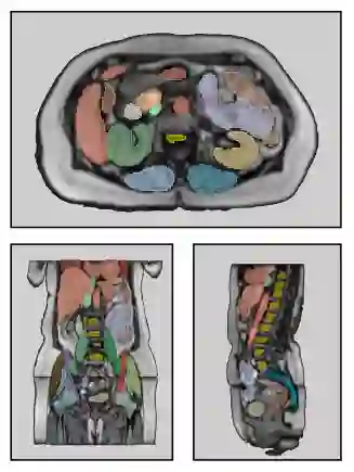
Purpose: To introduce a deep learning model capable of multi-organ segmentation in MRI scans, offering a solution to the current limitations in MRI analysis due to challenges in resolution, standardized intensity values, and variability in sequences. Materials and Methods: he model was trained on 1,200 manually annotated MRI scans from the UK Biobank, 221 in-house MRI scans and 1228 CT scans, leveraging cross-modality transfer learning from CT segmentation models. A human-in-the-loop annotation workflow was employed to efficiently create high-quality segmentations. The model's performance was evaluated on NAKO and the AMOS22 dataset containing 600 and 60 MRI examinations. Dice Similarity Coefficient (DSC) and Hausdorff Distance (HD) was used to assess segmentation accuracy. The model will be open sourced. Results: The model showcased high accuracy in segmenting well-defined organs, achieving Dice Similarity Coefficient (DSC) scores of 0.97 for the right and left lungs, and 0.95 for the heart. It also demonstrated robustness in organs like the liver (DSC: 0.96) and kidneys (DSC: 0.95 left, 0.95 right), which present more variability. However, segmentation of smaller and complex structures such as the portal and splenic veins (DSC: 0.54) and adrenal glands (DSC: 0.65 left, 0.61 right) revealed the need for further model optimization. Conclusion: The proposed model is a robust, tool for accurate segmentation of 40 anatomical structures in MRI and CT images. By leveraging cross-modality learning and interactive annotation, the model achieves strong performance and generalizability across diverse datasets, making it a valuable resource for researchers and clinicians. It is open source and can be downloaded from https://github.com/hhaentze/MRSegmentator.
翻译:目的:引入一种能够在MRI扫描中实现多器官分割的深度学习模型,以解决当前MRI分析因分辨率、标准化强度值及序列变异性挑战而存在的局限性。材料与方法:该模型基于英国生物银行(UK Biobank)的1,200例手动标注MRI扫描、221例内部MRI扫描及1,228例CT扫描进行训练,利用跨模态迁移学习技术从CT分割模型中迁移知识。采用人机协同标注工作流程高效生成高质量分割结果。模型性能在包含600例和60例MRI检查的NAKO数据集与AMOS22数据集上进行了评估,使用Dice相似系数(DSC)和豪斯多夫距离(HD)量化分割精度。该模型将开源发布。结果:模型在分割边界清晰的器官时展现出高精度,右肺与左肺的Dice相似系数(DSC)达0.97,心脏达0.95。对于肝脏(DSC:0.96)和肾脏(左肾DSC:0.95,右肾DSC:0.95)等变异性较大的器官,模型同样表现出鲁棒性。然而,门静脉与脾静脉(DSC:0.54)及肾上腺(左侧DSC:0.65,右侧DSC:0.61)等较小或复杂结构的分割结果表明仍需进一步优化模型。结论:所提模型是一种能够对MRI与CT图像中40种解剖结构进行精确分割的鲁棒工具。通过结合跨模态学习与交互式标注,模型实现了优异性能并在多样化数据集中具备良好泛化能力,为研究人员和临床医生提供了宝贵资源。模型已开源,可从https://github.com/hhaentze/MRSegmentator下载。






相关内容




 微信扫码咨询专知VIP会员