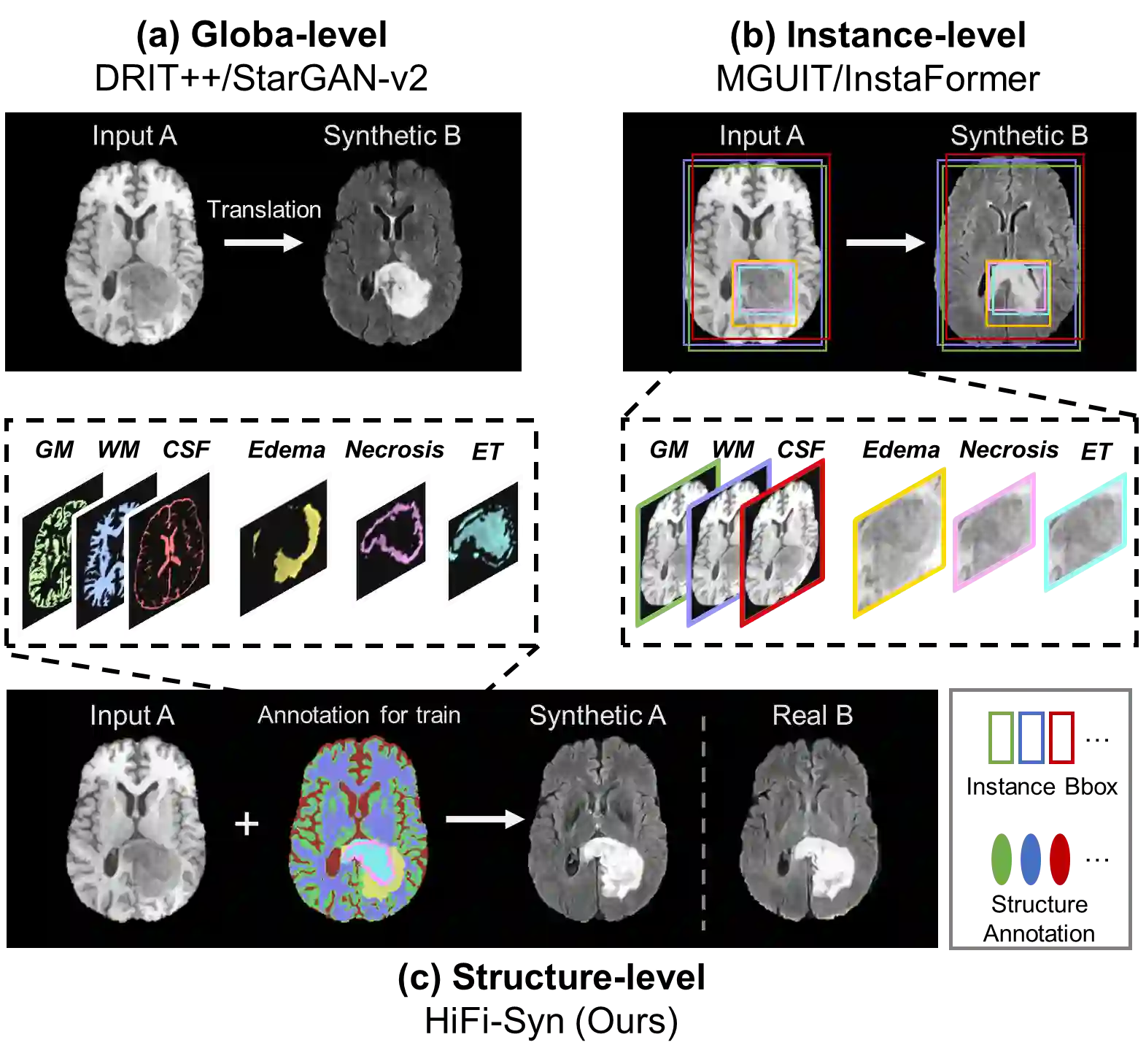
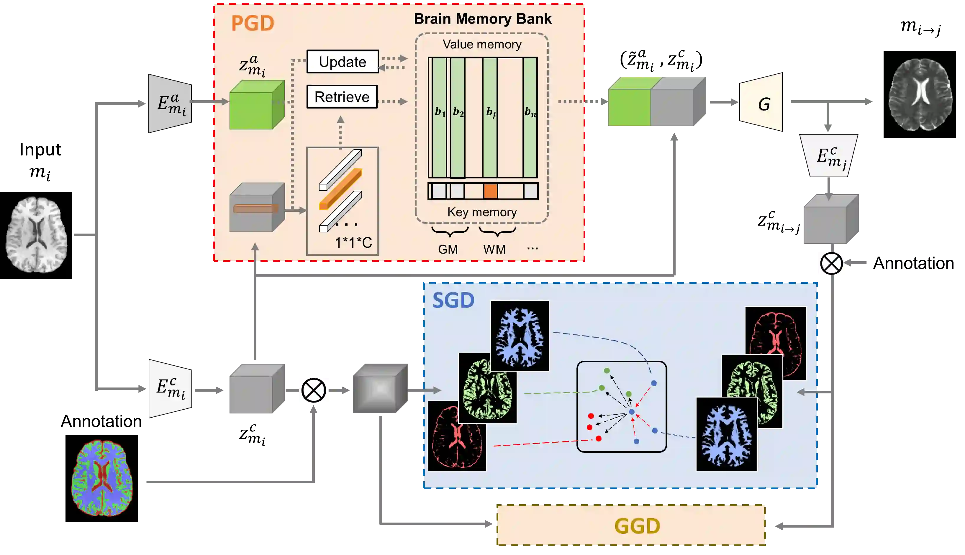
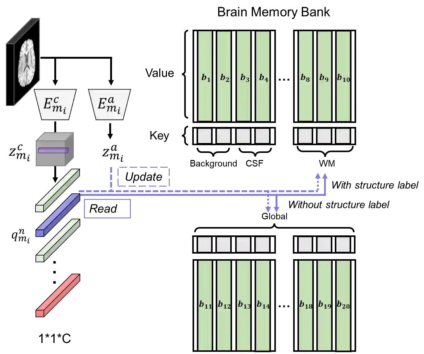
Synthesizing medical images while preserving their structural information is crucial in medical research. In such scenarios, the preservation of anatomical content becomes especially important. Although recent advances have been made by incorporating instance-level information to guide translation, these methods overlook the spatial coherence of structural-level representation and the anatomical invariance of content during translation. To address these issues, we introduce hierarchical granularity discrimination, which exploits various levels of semantic information present in medical images. Our strategy utilizes three levels of discrimination granularity: pixel-level discrimination using a Brain Memory Bank, structure-level discrimination on each brain structure with a re-weighting strategy to focus on hard samples, and global-level discrimination to ensure anatomical consistency during translation. The image translation performance of our strategy has been evaluated on three independent datasets (UK Biobank, IXI, and BraTS 2018), and it has outperformed state-of-the-art algorithms. Particularly, our model excels not only in synthesizing normal structures but also in handling abnormal (pathological) structures, such as brain tumors, despite the variations in contrast observed across different imaging modalities due to their pathological characteristics. The diagnostic value of synthesized MR images containing brain tumors has been evaluated by radiologists. This indicates that our model may offer an alternative solution in scenarios where specific MR modalities of patients are unavailable. Extensive experiments further demonstrate the versatility of our method, providing unique insights into medical image translation.
翻译:在医学图像合成中保持其结构信息对于医学研究至关重要。在此类场景下,解剖内容的保持尤为重要。尽管近期研究通过引入实例级信息来指导图像转换取得了进展,但这些方法忽视了结构级表示的空间连贯性以及转换过程中内容的解剖不变性。为解决这些问题,我们提出了层次粒度判别方法,该方法充分利用了医学图像中存在的多层级语义信息。我们的策略采用三个层次的判别粒度:利用脑记忆库进行像素级判别;对每个脑结构采用重加权策略进行结构级判别以聚焦于困难样本;以及全局级判别以确保转换过程中的解剖一致性。我们的图像转换策略已在三个独立数据集(UK Biobank、IXI 和 BraTS 2018)上进行了性能评估,并超越了现有最先进算法。值得注意的是,尽管不同成像模态因病理特性而呈现对比度差异,我们的模型不仅在合成正常结构方面表现优异,还能有效处理异常(病理)结构(如脑肿瘤)。包含脑肿瘤的合成磁共振图像的诊断价值已由放射科医生进行评估。这表明在患者特定磁共振模态不可用的情况下,我们的模型可能提供一种替代解决方案。大量实验进一步证明了我们方法的通用性,为医学图像转换领域提供了独特见解。