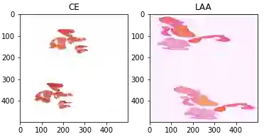
An innovative two-stage methodology for categorizing blood clot origins is presented in this paper, which is important for the diagnosis and treatment of ischemic stroke. First, a background classifier based on MobileNetV3 segments big whole-slide digital pathology images into numerous tiles to detect the presence of cellular material. After that, different pre-trained image classification algorithms are fine-tuned to determine the origin of blood clots. Due to complex blood flow dynamics and limitations in conventional imaging methods such as computed tomography (CT), magnetic resonance imaging (MRI), and ultrasound, identifying the sources of blood clots is a challenging task. Although these techniques are useful for identifying blood clots, they are not very good at determining how they originated. To address these challenges, our method makes use of robust computer vision models that have been refined using information from whole-slide digital pathology images. Out of all the models tested, the PoolFormer \cite{yu2022metaformer} performs better than the others, with 93.4\% accuracy, 93.4\% precision, 93.4\% recall, and 93.4\% F1-score. Moreover, it achieves the good weighted multi-class logarithmic loss (WMCLL) of 0.4361, which emphasizes how effective it is in this particular application. These encouraging findings suggest that our approach can successfully identify the origin of blood clots in a variety of vascular locations, potentially advancing ischemic stroke diagnosis and treatment approaches.
翻译:本文提出了一种创新的两阶段方法,用于对血凝块起源进行分类,这对缺血性卒中的诊断和治疗具有重要意义。首先,基于MobileNetV3的背景分类器将大型全切片数字病理图像分割为多个图块,以检测细胞物质的存在。随后,对不同的预训练图像分类算法进行微调,以确定血凝块的起源。由于复杂的血流动力学以及计算机断层扫描(CT)、磁共振成像(MRI)和超声等传统成像方法的局限性,识别血凝块来源是一项具有挑战性的任务。尽管这些技术对识别血凝块有用,但它们并不擅长确定其来源。为应对这些挑战,我们的方法利用了经过全切片数字病理图像数据优化的鲁棒计算机视觉模型。在所有测试的模型中,PoolFormer \cite{yu2022metaformer} 的表现优于其他模型,准确率达93.4%,精确率达93.4%,召回率达93.4%,F1分数达93.4%。此外,它还取得了良好的加权多类对数损失(WMCLL)0.4361,这突显了其在此特定应用中的有效性。这些令人鼓舞的结果表明,我们的方法能够成功识别不同血管位置的血凝块起源,有望推进缺血性卒中的诊断和治疗方法。