

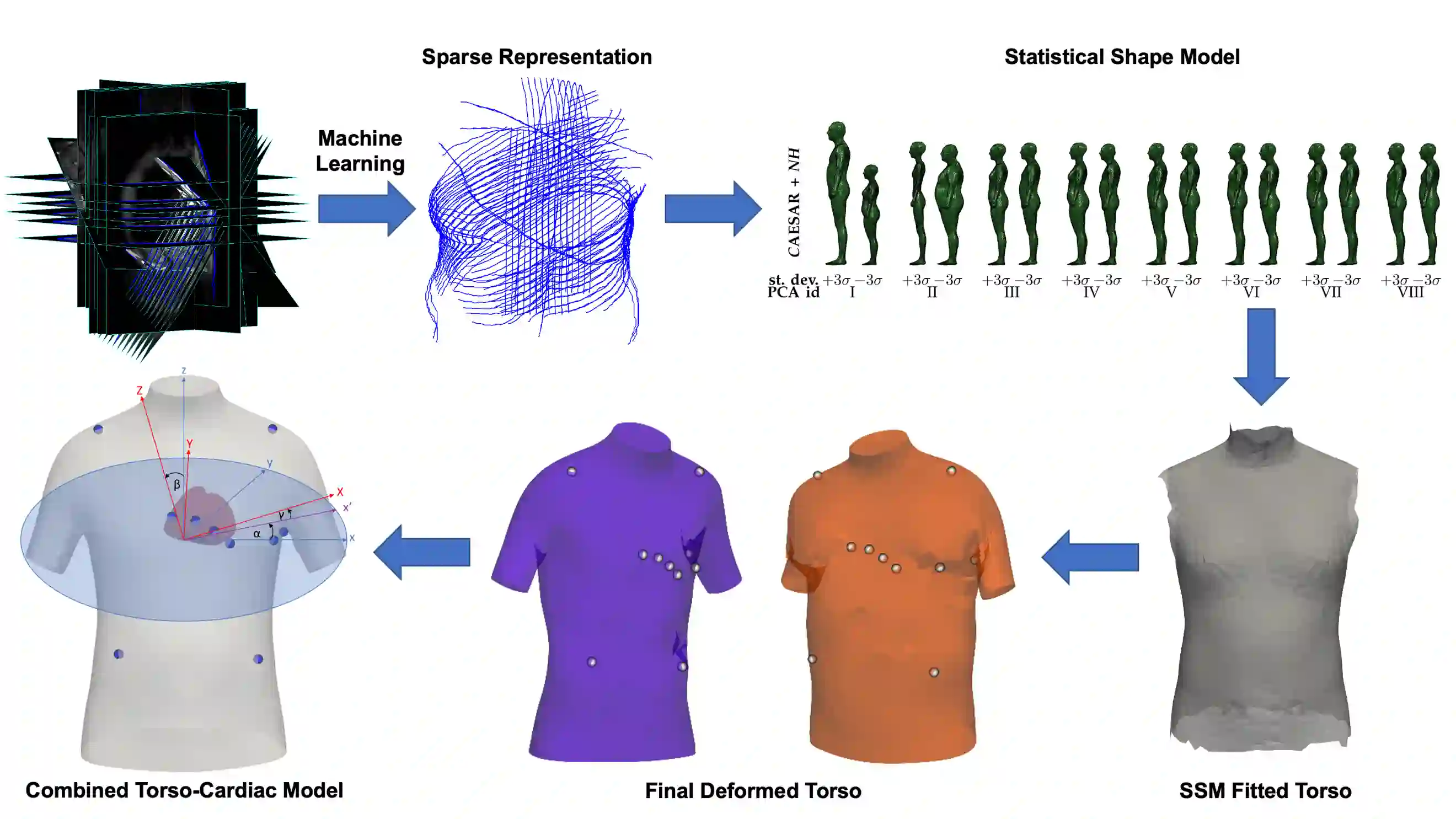
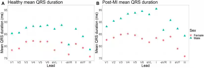
The electrocardiogram (ECG) is routinely used in cardiology, though its interpretation is confounded by anatomical variability. A novel, automated computational pipeline enables quantification of torso-ventricular anatomy metrics from magnetic resonance imaging, and comparison to ECG characteristics. Sex and myocardial infarction differences are investigated based on 1051 healthy and 425 post-MI subjects from UK Biobank. Smaller ventricles in females explain ~50% of shorter QRS durations than in males, and contribute to lower STJ amplitudes in females (also due to more superior and posterior position). In females, torso-ventricular anatomy, particularly from larger BMI, is a stronger modulator of T wave amplitude reductions and left-deviated R axis angles in post-MI than in males. Thus, female MI phenotype is less reflective of pathology, and baseline STJ amplitudes and QRS durations are further from clinical thresholds. Therefore, quantification of anatomical sex-differences and impact on ECG in health and disease is critical to avoid clinical sex-bias.
翻译:心电图是心脏病学中常规使用的诊断工具,但其解读常受解剖变异性的干扰。本研究提出了一种新型自动化计算流程,可从磁共振成像中量化躯干-心室解剖指标,并与心电图特征进行对比分析。基于英国生物银行1051名健康受试者和425名心肌梗死后受试者的数据,我们探究了性别差异及心肌梗死的影响。女性心室体积较小可解释其QRS波群时程较男性缩短约50%的原因,并导致STJ振幅降低(亦因心脏位置更偏上偏后)。在女性心肌梗死后患者中,躯干-心室解剖(尤其由较大体重指数所致)对T波振幅降低和R电轴左偏的调节作用强于男性。因此,女性心肌梗死的电表型对病理状态的反映较弱,且基线STJ振幅与QRS时程更远离临床阈值。准确定量化解剖学性别差异及其对健康与疾病状态下心电图的影响,对于避免临床性别偏倚至关重要。






相关内容




 微信扫码咨询专知VIP会员