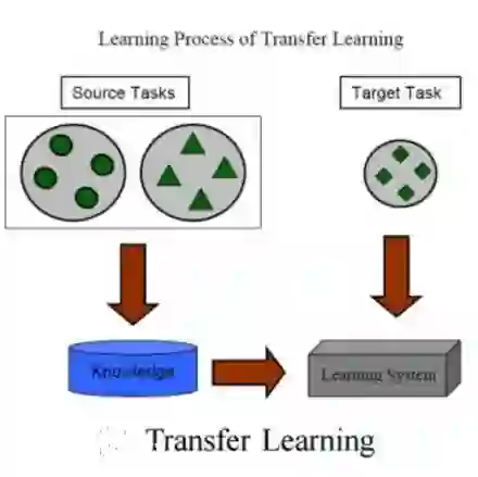
The accurate understanding of ischemic stroke lesions is critical for efficient therapy and prognosis of stroke patients. Magnetic resonance imaging (MRI) is sensitive to acute ischemic stroke and is a common diagnostic method for stroke. However, manual lesion segmentation performed by experts is tedious, time-consuming, and prone to observer inconsistency. Automatic medical image analysis methods have been proposed to overcome this challenge. However, previous approaches have relied on hand-crafted features that may not capture the irregular and physiologically complex shapes of ischemic stroke lesions. In this study, we present a novel framework for quickly and automatically segmenting ischemic stroke lesions on various MRI sequences, including T1-weighted, T2-weighted, DWI, and FLAIR. The proposed methodology is validated on the ISLES 2015 Brain Stroke sequence dataset, where we trained our model using the Res-Unet architecture twice: first, with pre-existing weights, and then without, to explore the benefits of transfer learning. Evaluation metrics, including the Dice score and sensitivity, were computed across 3D volumes. Finally, a Majority Voting Classifier was integrated to amalgamate the outcomes from each axis, resulting in a comprehensive segmentation method. Our efforts culminated in achieving a Dice score of 80.5\% and an accuracy of 74.03\%, showcasing the efficacy of our segmentation approach.
翻译:准确理解缺血性脑卒中病灶对于卒中患者的有效治疗和预后评估至关重要。磁共振成像(MRI)对急性缺血性脑卒中具有高敏感性,是脑卒中的常用诊断方法。然而,由专家进行的手动病灶分割过程繁琐、耗时,且易受观察者间差异影响。为应对这一挑战,已有自动医学图像分析方法被提出。然而,既往方法依赖于手工设计的特征,可能无法捕捉缺血性脑卒中病灶不规则且生理结构复杂的形态。本研究提出一种新颖框架,可在多种MRI序列(包括T1加权、T2加权、弥散加权成像及液体衰减反转恢复序列)上快速自动分割缺血性脑卒中病灶。所提方法在ISLES 2015脑卒中序列数据集上得到验证:我们采用Res-Unet架构进行两次模型训练——首次使用预训练权重,随后不使用权重,以探究迁移学习的优势。通过三维体积数据计算了戴斯相似系数及敏感度等评估指标。最终集成多数投票分类器融合各轴向分割结果,形成综合分割方案。本研究最终实现80.5%的戴斯系数与74.03%的准确率,充分证明了所提分割方法的有效性。