

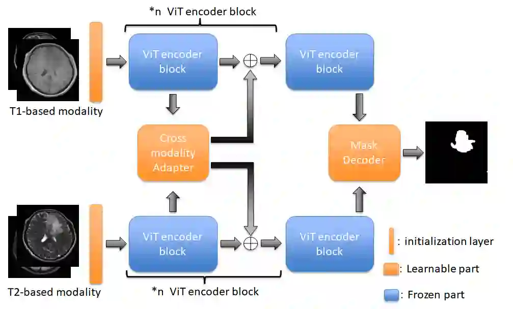
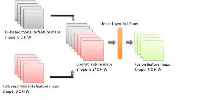
According to the 2021 World Health Organization (WHO) Classification scheme for gliomas, glioma segmentation is a very important basis for diagnosis and genotype prediction. In general, 3D multimodal brain MRI is an effective diagnostic tool. In the past decade, there has been an increase in the use of machine learning, particularly deep learning, for medical images processing. Thanks to the development of foundation models, models pre-trained with large-scale datasets have achieved better results on a variety of tasks. However, for medical images with small dataset sizes, deep learning methods struggle to achieve better results on real-world image datasets. In this paper, we propose a cross-modality attention adapter based on multimodal fusion to fine-tune the foundation model to accomplish the task of glioma segmentation in multimodal MRI brain images with better results. The effectiveness of the proposed method is validated via our private glioma data set from the First Affiliated Hospital of Zhengzhou University (FHZU) in Zhengzhou, China. Our proposed method is superior to current state-of-the-art methods with a Dice of 88.38% and Hausdorff distance of 10.64, thereby exhibiting a 4% increase in Dice to segment the glioma region for glioma treatment.
翻译:根据2021年世界卫生组织(WHO)胶质瘤分类方案,胶质瘤分割是诊断和基因型预测的重要依据。通常,三维多模态脑部MRI是一种有效的诊断工具。过去十年中,机器学习(尤其是深度学习)在医学图像处理中的应用日益增多。得益于基础模型的发展,通过大规模数据集预训练的模型在多种任务中取得了更优的结果。然而,对于数据集规模较小的医学图像,深度学习方法难以在真实图像数据集上获得更好的效果。本文提出一种基于多模态融合的跨模态注意力适配器,用于微调基础模型,从而在多模态MRI脑部图像中更好地完成胶质瘤分割任务。通过来自中国郑州大学第一附属医院(FHZU)的私有胶质瘤数据集验证了所提方法的有效性。我们的方法优于当前最先进的方法,Dice系数达到88.38%,豪斯多夫距离为10.64,在分割胶质瘤区域以实现胶质瘤治疗方面,Dice系数提升了4%。





相关内容



 微信扫码咨询专知VIP会员