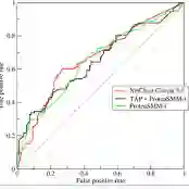
Purpose: To develop a method for automated segmentation of hypothalamus subregions informed by ultra-high resolution ex vivo magnetic resonance images (MRI), which generalizes across MRI sequences and resolutions without retraining. Materials and Methods: We trained our deep learning method, H-synEx, with synthetic images derived from label maps built from ultra-high resolution ex vivo MRI scans, which enables finer-grained manual segmentation when compared with 1mm isometric in vivo images. We validated this retrospective study using 1535 in vivo images from six datasets and six MRI sequences. The quantitative evaluation used the Dice Coefficient (DC) and Average Hausdorff distance (AVD). Statistical analysis compared hypothalamic subregion volumes in controls, Alzheimer's disease (AD), and behavioral variant frontotemporal dementia (bvFTD) subjects using the area under the curve (AUC) and Wilcoxon rank sum test. Results: H-SynEx can segment the hypothalamus across various MRI sequences, encompassing FLAIR sequences with significant slice spacing (5mm). Using hypothalamic volumes on T1w images to distinguish control from AD and bvFTD patients, we observed AUC values of 0.74 and 0.79 respectively. Additionally, AUC=0.66 was found for volume variation on FLAIR scans when comparing control and non-patients. Conclusion: Our results show that H-SynEx successfully leverages information from ultra-high resolution scans to segment in vivo from different MRI sequences such as T1w, T2w, PD, qT1, FA, and FLAIR. We also found that our automated segmentation was able to discriminate controls versus patients on FLAIR images with 5mm spacing. H-SynEx is openly available at https://github.com/liviamarodrigues/hsynex.
翻译:摘要:目的:开发一种基于超高分辨率离体磁共振图像(MRI)的下丘脑亚区自动分割方法,该方法无需重新训练即可泛化至不同MRI序列和分辨率。材料与方法:我们训练的深度学习方法H-SynEx,利用由超高分辨率离体MRI扫描生成的标签图构建合成图像,相较于1mm等距活体图像,实现了更精细的手动分割。本研究回顾性验证使用来自六个数据集和六种MRI序列的1535幅活体图像。定量评估采用Dice系数(DC)与平均豪斯多夫距离(AVD)。统计学分析通过曲线下面积(AUC)和威尔科克森秩和检验,比较对照组、阿尔茨海默病(AD)及行为变异型额颞叶痴呆(bvFTD)患者的下丘脑亚区体积。结果:H-SynEx可分割多种MRI序列下的下丘脑,包括层间距显著(5mm)的FLAIR序列。利用T1加权图像上的下丘脑体积区分对照组与AD及bvFTD患者时,我们观察到AUC值分别为0.74和0.79。此外,在FLAIR扫描中比较对照组与非患者时,体积变化的AUC=0.66。结论:我们的结果表明H-SynEx成功利用超高分辨率扫描信息分割来自不同MRI序列(如T1w、T2w、PD、qT1、FA和FLAIR)的活体图像。我们还发现,自动分割能区分5mm层间距FLAIR图像中的对照组与患者。H-SynEx已开源发布于https://github.com/liviamarodrigues/hsynex。