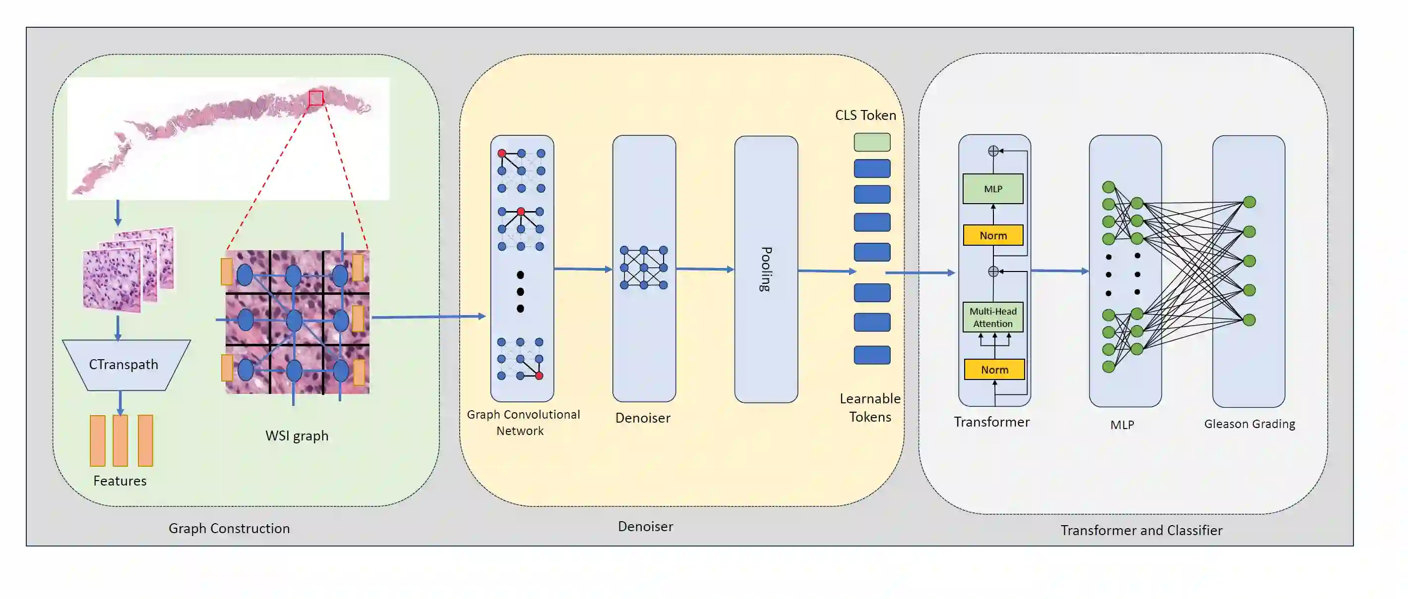
Whole slide images~(WSIs) are digitized images of tissues placed in glass slides using advanced scanners. The digital processing of WSIs is challenging as they are gigapixel images and stored in multi-resolution format. A common challenge with WSIs is that perturbations/artifacts are inevitable during storing the glass slides and digitizing them. These perturbations include motion, which often arises from slide movement during placement, and changes in hue and brightness due to variations in staining chemicals and the quality of digitizing scanners. In this work, a novel robust learning approach to account for these artifacts is presented. Due to the size and resolution of WSIs and to account for neighborhood information, graph-based methods are called for. We use graph convolutional network~(GCN) to extract features from the graph representing WSI. Through a denoiser {and pooling layer}, the effects of perturbations in WSIs are controlled and the output is followed by a transformer for the classification of different grades of prostate cancer. To compare the efficacy of the proposed approach, the model without denoiser is trained and tested with WSIs without any perturbation and then different perturbations are introduced in WSIs and passed through the network with the denoiser. The accuracy and kappa scores of the proposed model with prostate cancer dataset compared with non-robust algorithms show significant improvement in cancer diagnosis.
翻译:全切片图像(WSI)是利用先进扫描仪对置于玻片中的组织进行数字化处理得到的图像。由于WSI为千兆像素级图像并以多分辨率格式存储,其数字处理面临诸多挑战。WSI的一个常见难题是:在玻片存储与数字化过程中,扰动/伪影难以避免。这些扰动包括因玻片放置时移动产生的运动伪影,以及因染色剂差异和扫描仪质量变化导致的色相与亮度波动。本研究提出了一种针对此类伪影的新型鲁棒学习方法。考虑到WSI的尺寸与分辨率特性,并兼顾邻域信息需求,我们采用基于图的方法。具体而言,利用图卷积网络(GCN)从表征WSI的图中提取特征。通过去噪层与池化层的协同作用,有效控制WSI中扰动的影响,随后将输出输入Transformer以完成前列腺癌不同等级的分类任务。为验证所提方法的有效性,我们在无扰动WSI上训练并测试了不含去噪层的基准模型,随后在WSI中引入不同扰动并通过含去噪层的网络进行处理。基于前列腺癌数据集的实验结果表明,相较非鲁棒算法,本模型的准确率与Kappa系数在前列腺癌诊断方面均取得显著提升。