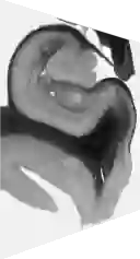
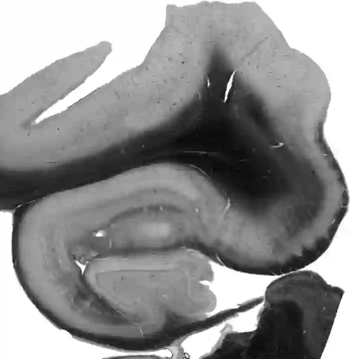
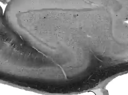
Understanding the cortical organization of the human brain requires interpretable descriptors for distinct structural and functional imaging data. 3D polarized light imaging (3D-PLI) is an imaging modality for visualizing fiber architecture in postmortem brains with high resolution that also captures the presence of cell bodies, for example, to identify hippocampal subfields. The rich texture in 3D-PLI images, however, makes this modality particularly difficult to analyze and best practices for characterizing architectonic patterns still need to be established. In this work, we demonstrate a novel method to analyze the regional organization of the human hippocampus in 3D-PLI by combining recent advances in unfolding methods with deep texture features obtained using a self-supervised contrastive learning approach. We identify clusters in the representations that correspond well with classical descriptions of hippocampal subfields, lending validity to the developed methodology.
翻译:理解人类大脑皮层组织需要针对不同结构和功能影像数据构建可解释的描述符。三维偏振光成像(3D-PLI)是一种高分辨率可视化死后脑组织纤维结构的成像模态,同时能捕捉细胞体存在信息,例如用于识别海马亚区。然而,3D-PLI图像中丰富的纹理特征使该模态的分析极具挑战性,目前仍需建立表征结构模式的规范方法。本研究通过将自监督对比学习获得的深度纹理特征与展开方法的最新进展相结合,提出了一种分析人类海马体在3D-PLI中区域组织的新方法。我们在特征表示中识别出的聚类与经典海马亚区描述高度吻合,验证了所开发方法的有效性。