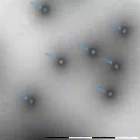
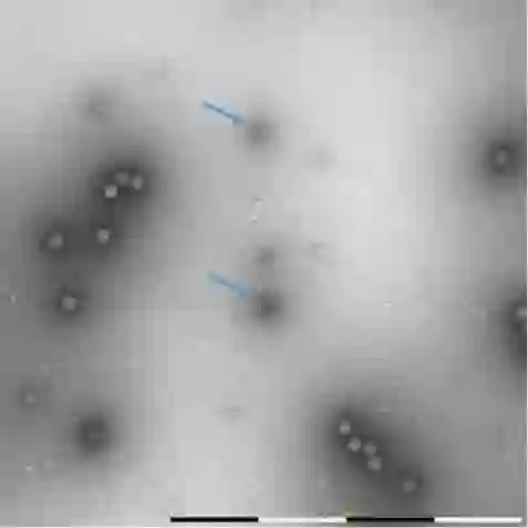
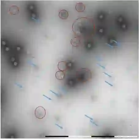
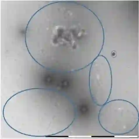
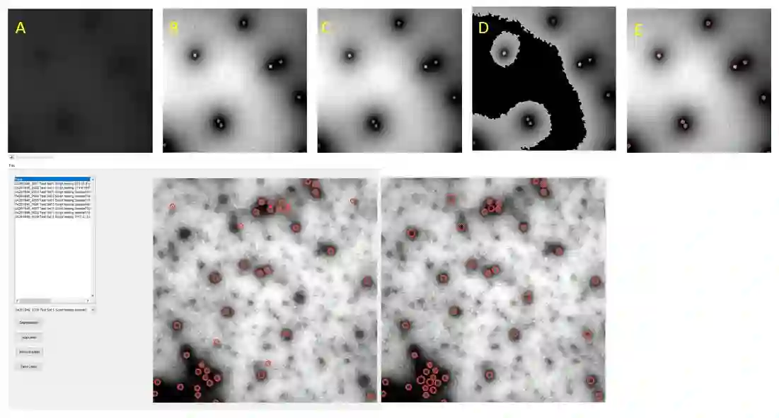
Regular monitoring of the primary particles and purity profiles of a drug product during development and manufacturing processes is essential for manufacturers to avoid product variability and contamination. Transmission electron microscopy (TEM) imaging helps manufacturers predict how changes affect particle characteristics and purity for virus-based gene therapy vector products and intermediates. Since intact particles can characterize efficacious products, it is beneficial to automate the detection of intact adenovirus against a non-intact-viral background mixed with debris, broken, and artefact particles. In the presence of such particles, detecting intact adenoviruses becomes more challenging. To overcome the challenge, due to such a presence, we developed a software tool for semi-automatic annotation and segmentation of adenoviruses and a software tool for automatic segmentation and detection of intact adenoviruses in TEM imaging systems. The developed semi-automatic tool exploited conventional image analysis techniques while the automatic tool was built based on convolutional neural networks and image analysis techniques. Our quantitative and qualitative evaluations showed outstanding true positive detection rates compared to false positive and negative rates where adenoviruses were nicely detected without mistaking them for real debris, broken adenoviruses, and/or staining artefacts.
翻译:在药品开发和制造过程中,定期监测活性颗粒及药品纯度曲线对于制造商避免产品变异和污染至关重要。透射电镜成像技术可帮助制造商预测变化如何影响基于病毒的基因治疗载体产品及中间体的颗粒特性和纯度。由于完整颗粒能够表征有效产品,因此在混有碎片、破损颗粒和假象的非完整病毒背景中自动检测完整腺病毒具有重要意义。当存在此类颗粒时,检测完整腺病毒变得更具挑战性。为克服这一挑战,我们开发了用于腺病毒半自动标注和分割的软件工具,以及用于透射电镜成像系统中完整腺病毒自动分割和检测的软件工具。所开发的半自动工具采用传统图像分析技术,而自动工具基于卷积神经网络与图像分析技术构建。定量和定性评估显示,与假阳性率和假阴性率相比,我们的真阳性检测率表现优异,腺病毒被准确检出而未与真实碎片、破损腺病毒或染色假象混淆。